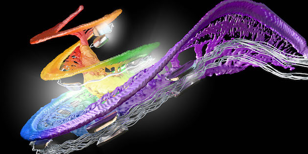
THE COCHLEA AS YOU’VE NEVER SEEN IT BEFORE
MED-EL’s deal to share academics’ “unimaginable” anatomy dataset brings what the firm’s leader hails as a “new era” in cochlear implants.

Austrian Cochlear implant manufacturer MED-EL has signed a new agreement with Western University (London, Canada) and the non-profit Canadian research organisation Mitacs for exclusive access to an extraordinary synchrotron dataset of temporal bones, one that brings the implant pioneers an unprecedented level of details of human (inner) ears, reliably revealing their individual variations.
MED-EL agreed to an exclusive share in Audiology News UK of the images accompanying this text, no mere set of useful slides, the company points out, but a groundbreaking collection that offers the highest resolution and most comprehensive view of cochlear anatomy to date.
It will give MED-EL opportunities to rethink and develop innovative pre-operative decision-making processes, personalised and precise cochlear implantation procedures, enhancement of existing cochlear implantation solutions, as well as fitting processes tailored to the capabilities and needs of individual cochlear implant users.
Furthermore, the company underlines, translating these goals across products and services falls in line with MED-EL’s focus on hearing preservation and enabling the closest to natural hearing including music perception and speech understanding for users.
What is synchrotron imaging?
The intense beams of light produced by a synchroton particle accelerator allow for special views of mechanisms in medical and many other areas of research.
For MED-EL, synchrotron images provide incredible detail, revealing individual variations in the length and shape of the inner ear.
“This agreement marks the start of a new era in the field of cochlear implants,” proclaims Ingeborg Hochmair, CEO of MED-EL. “We can now take into account candidates’ individual inner ear specifics and anatomical characteristics to an extent that was previously unimaginable.”
“By harnessing this cutting-edge dataset, we are now equipped with the tools and insights to revolutionise the entire process of cochlear implantation, from pre-operative planning to surgery and fitting,” adds Hochmair.
The deal is articulated around a collaboration between MED-EL and Principal Investigators Sumit Agrawal and Hanif Ladak at Western University. This partnership brings the CI pioneer firm the academics’ AI-based technology and software code, coupled with terabytes of image data. “For cochlear implant recipients, this represents an unprecedented opportunity to achieve a new level of natural hearing,” says the firm.
SCALA WITH COLOURED OVERLAYS
A cross-sectional image of the cochlea showing the scala tympani, scala media and scala vestibuli, emphasising with coloured overlays parts of the basilar membrane and their associated neural projections. In the centre of the image is the modiolus.
VESTIBULAR ORGAN AND COCHLEA
A 3D Synchrotron image of the vestibular organ and cochlea includes an overlay to show tonotopically aligned frequency ranges along the basilar membrane.
OSSICLES
This rendered view of the cochlea provides tonotopically aligned frequency ranges along the basilar membrane, whilst also drawing attention to the neural structures of the cochlea from the basal to the apical region. This 3D image also provides a clear view of the ossicles including malleus (partly hiding the stapes), incus, stapes, stapes footplate covering the oval window, as well the vestibular organ.
CI ELECTRODE ARRAY
A projection view of the cochlea highlighting some of its delicate structures and showcasing an inserted MED-EL FlexSoft cochlear implant electrode array which has been colour-illustrated to highlight tonotopically-aligned frequency ranges.
COCHLEAR HOOK REGION EXPLODED
A 3D Synchrotron image providing precision details of the basilar membrane and neural structures from the hook region all the way to the apex of the cochlea, overlaid by electrical contacts and wires of a fully inserted MED-EL FlexSoft cochlear implant electrode array.
CI ELECTRODE ARRAY WITH TONOTOPIC FREQUENCIES
The same image of the CI electrode array (above), additionally emphasising the relation of the inserted electrode contacts to their insertion depths and corresponding tonotopic frequencies.
SEMICIRCULAR CANALS
This synchrotron image provides a 3D view of one of the three semicircular canals of the vestibular organ together with the cochlea intersected by a 2-dimensional plane showing the surrounding of the cochlea.
BASILAR MEMBRANE AND NEURAL STRUCTURES
This rendered view of the cochlea emphasizes the delicate basilar membrane spanning from base to apex and projecting neural structures of the cochlea, all of them colour-coded to schematically highlight various tonotopic frequency regions. The auditory nerve is shown on the bottom of the image.
FACIAL NERVE, VESTIBULAR ORGAN, ROUND WINDOW
An intricate view of the facial nerve (light yellow), facial recess, chorda tympani (dark yellow), vestibular organ (grey), ossicles (white), the round window of the cochlea (dark red), and the cochlea.
Source: MED-EL


